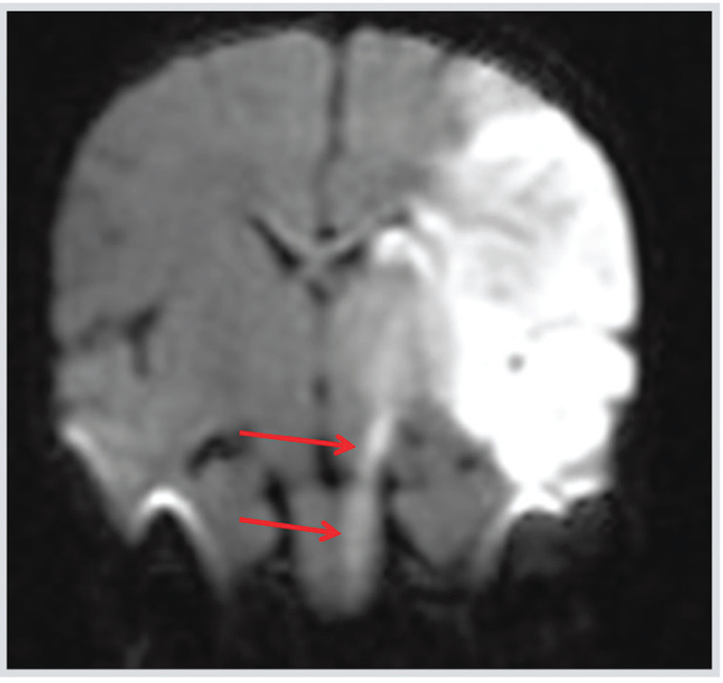
Figure 7-1.

Coronal diffusion-weighted image (DWI) of the patient in Case 7-1 showing a neonatal acute left middle cerebral artery arterial ischemic stroke showing acute Wallerian degeneration along the descending corticospinal tract from the infarcted area to the midbrain (cerebral peduncle) (arrows).
