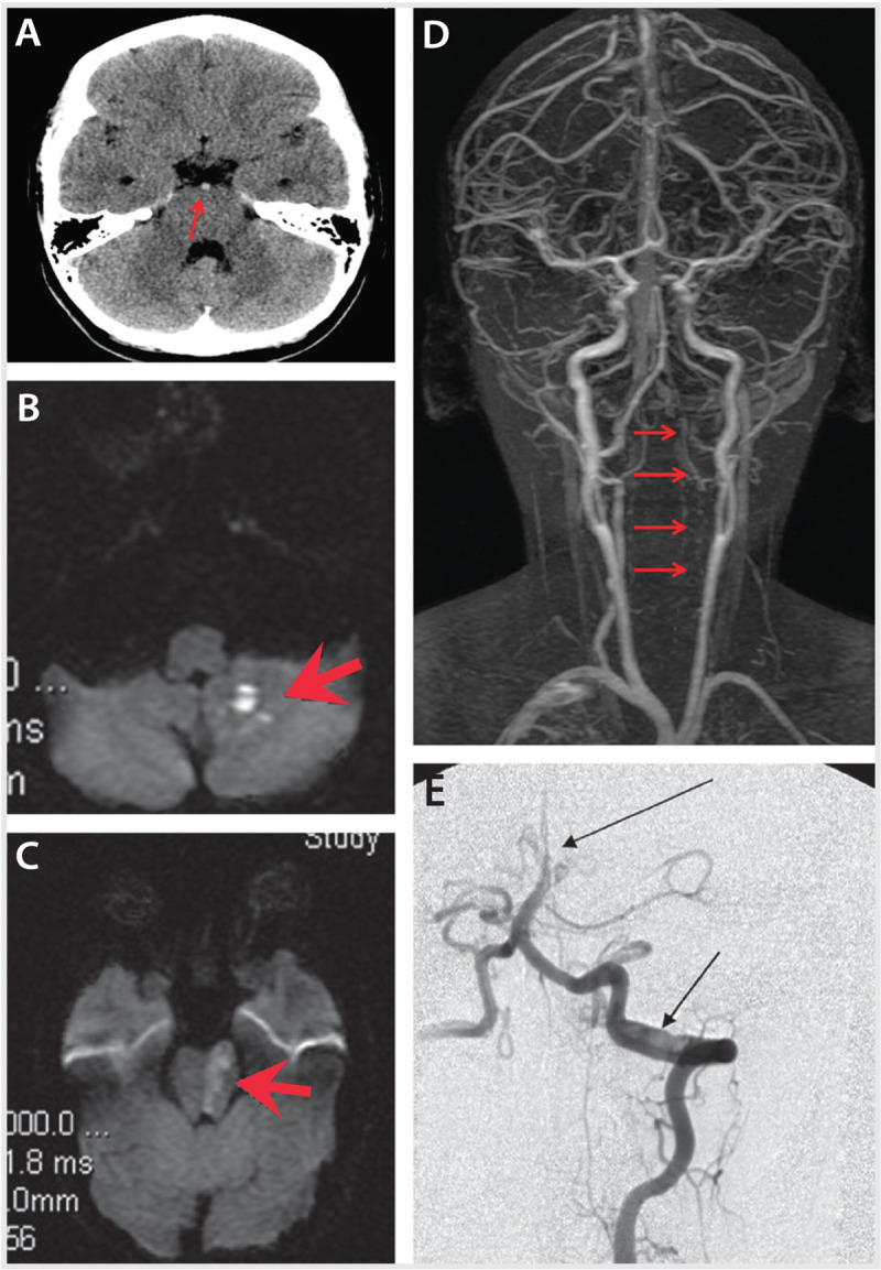
Figure 7-2.

Imaging studies at presentation of the patient in Case 7-2. A, Noncontrast head CT reveals hyperdense basilar artery (arrow). B and C, Diffusion-weighted image (DWI) reveals ischemia (arrows) in the left inferior cerebellar peduncle and left pons. D, Magnetic resonance angiography reveals greatly reduced flow in the entire left vertebral artery throughout its course (arrows). E, Catheter cerebral angiography reveals an intimal flap and double lumen (lower arrow) confirming left vertebral artery dissection at the C1-C2 vertebral level and occlusion of the distal one-third of the basilar artery (upper arrow).
