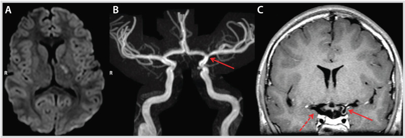
Figure 7-4.

Imaging studies at presentation of the patient in Case 7-3. Diffusion-weighted image (DWI) demonstrating diffusion restriction in the left internal capsule and globus pallidus (A), with narrowing of the distal left internal carotid artery (arrow) on magnetic resonance angiography (B). Arterial wall imaging (C) shows concentric wall enhancement of the distal left internal carotid artery, proximal middle cerebral artery, and proximal anterior cerebral artery (solid arrow) suggestive of inflammation compared with the opposite side (dashed arrow) that did not enhance.
