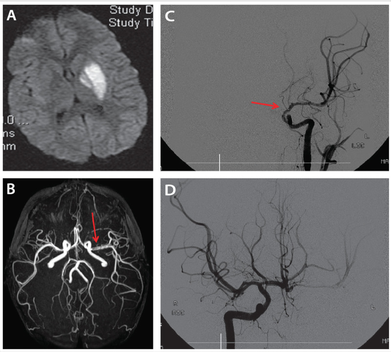
Figure 7-5.

A 4-year-old boy with transient cerebral arteriopathy. Diffusion-weighted image (DWI) (A) shows acute left globus pallidus and putamen infarct. Magnetic resonance angiography (B) shows irregular and reduced flow in the proximal left middle cerebral artery (arrow). Catheter cerebral angiography (C) reveals significant caliber reduction and irregularity (arrow) of the distal left internal carotid artery, proximal middle cerebral artery, and absent flow in the anterior cerebral artery with cross filling from the opposite side (D).
