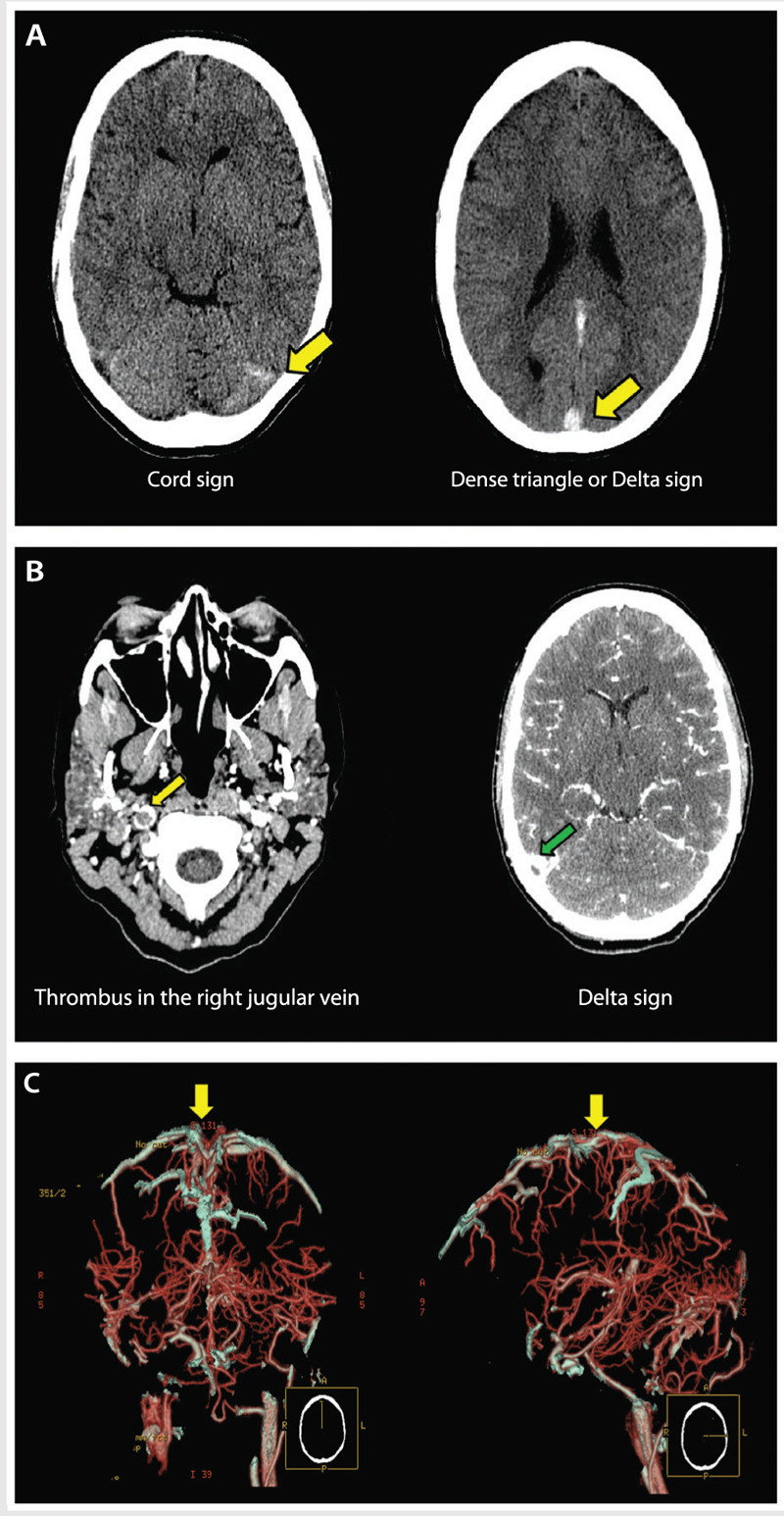
Figure 5-2.

A young woman presenting with headache in the emergency department. A, Plain CT shows increased attenuation involving the superior sagittal sinus, straight sinus, the confluence, and right transverse sinus (yellow arrows). B, CT venogram reveals absence of contrast in the superior sagittal sinus, straight sinus, right transverse sinus (green arrow), right sigmoid sinus, and right internal jugular vein (yellow arrow). C, Three-dimensional reconstruction of the CT venogram shows thrombosis of the superior sagittal sinus (yellow arrows).
