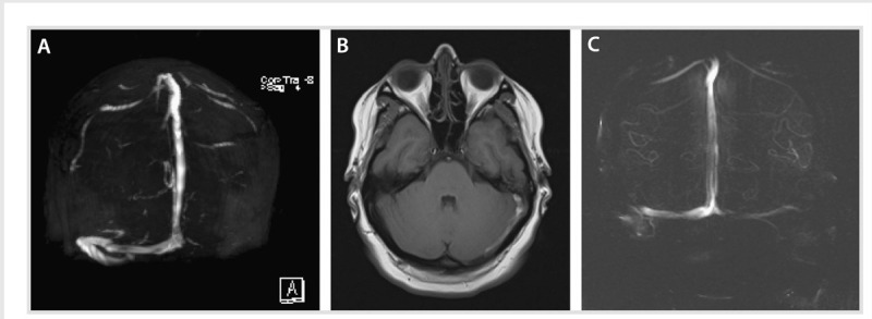
Figure 5-4.

A young pregnant woman with mastoiditis. A, Magnetic resonance venogram showing acute thrombosis of the left transverse and sigmoid sinuses. B, T1-weighted MRI of the brain showing left mastoid effusion and adjacent thrombosis that include the cortical veins of the tentorium. C, Resolution of the left transverse and sigmoid thrombosis 20 months later.
