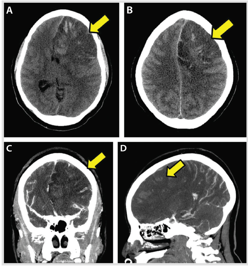
Figure 5-7.

Mass effect from the hemorrhagic conversion of a venous infarction (A–D, arrows) within the left frontal lobe lesion. Signs of brain herniation are evident, as is thrombosis of several left frontal cortical veins with very subtle extension in the superior sagittal sinus. The lack of visualization of residual lumen represents the occluded cortical veins and the associated severe mass effect from the adjacent brain edema (C, D, arrows).
