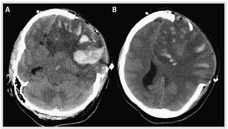
Figure 5-9.

Noncontrast CT post craniectomy showing further enlargement of the large left hemispheric lesion and associated multifocal hemorrhagic components and brain herniating through the craniectomy site with persistent signs of severe mass effect on the midline structures and basal cisterns. A, Inferior section, and B, superior section, both of which illustrate the mass effect and the hemorrhagic conversion.
