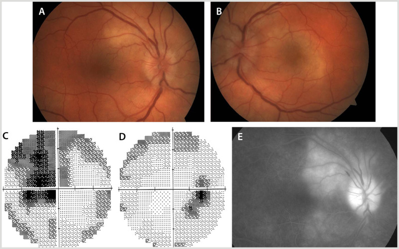
Figure 2-5.

A 58-year-old man with syphilis presented with vision loss in the left eye. Fundus examination showed bilateral optic disc edema (A, B). Visual field testing revealed a central visual field defect in the left eye (C) and an enlarged blind spot with an inferior temporal arcuate area of vision loss in the right eye (D). Fluorescein angiography of the right eye showed choroidal infiltrate around the optic disc (E).
Panels A, B, and C reprinted with permission from Almekhlafi MA, et al, Neurology.29 © 2011 AAN Enterprises, Inc. www.neurology.org/content/77/5/e28.long.
