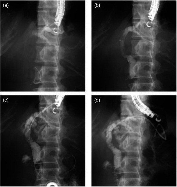
FIGURE 2.

(a) Following cholangiography, a 0.018‐inch guidewire was advanced and placed in the bile duct through a 22‐gauge FNA needle. (b) An ERCP catheter was inserted into the bile duct, bile was aspirated to decompress the bile duct, and cholangiography was performed to confirm the stenosis. (c) A laser‐cut type uncovered metal stent was deployed across the papilla. (d) A plastic stent was deployed from the intrahepatic bile duct to the jejunum.
