
Figure 1. The PLCG2M28L variant is associated with AD risk and downregulates PLCG2 expression.
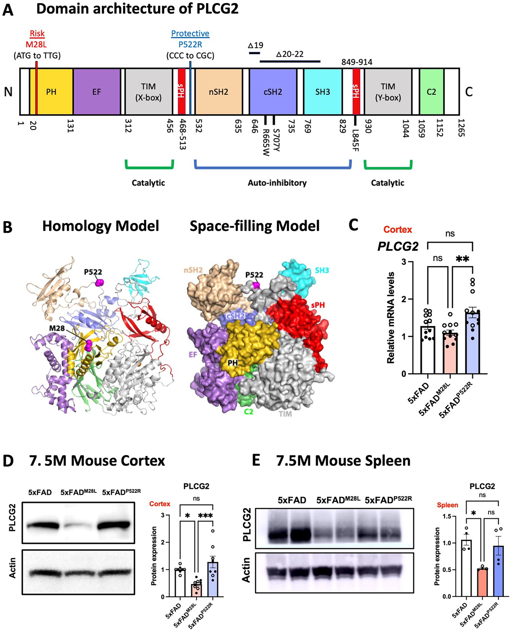
(A) Genetic linkage data of PLCG2M28L and PLCG2P522R with respect to AD risk are shown with the domain architecture of PLCG2 (to scale). Somatic mutations (R665W and S707Y) in PLCG2 are shown in the domain architecture. (B) PLCG2M28L (risk) and PLCG2P522R (protective) variants are mapped onto the structure of PLCG2 (magenta spheres) in both the homology model (left) and the space-filling model (right). (C). Gene expression of Plcg2 were assessed in cortical samples from 7.5-month-old 5xFAD, 5xFADM28L, and 5xFADP522R mice (n=12 per group; 6 male and 6 female mice). (D) Representative immunoblots and quantifications of PLCG2 protein expression in cortical lysates show reduced PLCG2 expression in 5xFADM28L mice (n=8 per group; 4 male and 4 female mice; 4 experiments). (E) Representative immunoblots and quantification of PLCG2 protein expression from the spleen show reduced PLCG2 expression in 5xFADM28L mice (n=4 per group; 2 male and 2 female mice; 2 experiments). All data are presented as the mean ± SEM, analyzed by an ordinary one-way ANOVA and Tukey’s multiple comparisons test. * P <0.05; ** P< 0.01; *** P< 0.001; ns: not significant. Male mice are marked with a solid circle (•), and the female mice are marked with a hollow circle (∘). See also Figure S1.
OR odds ratio, N amino-terminus, C carboxyl-terminus, PH pleckstrin homology domain, EF EF hand motif, TIM TIM barrel, sPH split PH domain, nSH2 n-terminus, Src Homology 2 domain, cSH2 c-terminus Src Homology 2 domain, SH3 SRC Homology 3 domain, C2 C2 domain, WT wild-type,