Fig. 1.
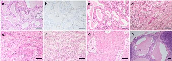
Histological and immunohistochemical features of LGASC with high-grade transformation (Case 1). a and b Histological features of core-needle biopsy. a The slide shows irregular dilation of the ducts with secretion in the lumen and moderate chronic inflammation in the stroma. b The immunohistochemical study showed that p40-positive cells existed discontinuously in the periphery of the ducts. c–h Histological findings of mastectomy specimens. c and d The superficial area. c The superficial area of the tumor consists of infiltrative glandular structures filled with abundant secretory materials in the lumen, resembling secretory carcinoma. The nuclear atypia of the tumor cells is bland. Few cells have mucin in their cytoplasm. d A view of a few tumor nests reveals squamous differentiation. e The deep area. The tumor displays clearer squamous cell differentiation. Some gland formations in the nests are seen. These findings are compatible with squamous cell carcinoma with the adenosquamous carcinoma pattern. f and g Histological findings of an axillary mass. f The lesions consisted of adenosquamous and spindle cell carcinoma. g Pure squamous cell carcinoma with a keratinization component is seen. h Some metastatic foci in the dissected lymph nodes include the LGASC component. Scale bar = 0.1 mm. LGASC, low-grade adenosquamous carcinoma

