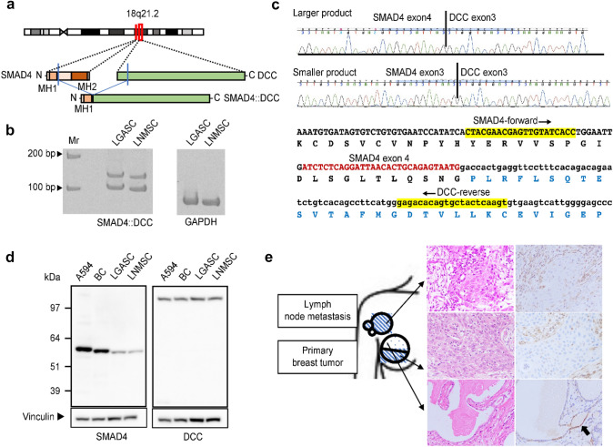
Fig. 5.
SMAD4::DCC fusion gene in LGASC and LNMSC. a The predicted SMAD4::DCC chimeric protein is schematically indicated. It is considered that the MH1 domain of SMAD4 is retained but that the MH2 domain of SMAD4 is lost. b RT‒PCR demonstrated the presence of two SMAD4::DCC fusion transcripts of different sizes in LGASC and LNMSC. On the right, two lanes show the internal glyceraldehyde 3-phosphate dehydrogenase control. Molecular weight markers are indicated at the left. The forward PCR primer targets exon 3 of SMAD4 (SMAD4-forward) and the reverse primer exon 3 of DCC (DCC-reverse). c Sanger sequencing revealed two chimeric transcripts of different sizes in both LGASC and LNMSC; the larger product is the SMAD4 exon 4–DCC exon 3 fusion transcript, and the smaller product is the SMAD4 exon 3–DCC exon 3 fusion transcript. d Western blotting with antibodies recognizing the N-terminus of SMAD4 (left) and the C-terminus of DCC (right) confirmed the existence of apparently wild-type SMAD4 and apparently wild-type DCC but not a protein derived from the SMAD4::DCC fusion. Molecular weight markers are indicated at the left. Vinculin as the loading control is shown at the bottom. On the left, two lanes show the positive controls (A594, lung cancer cell line; BC, breast cancer tissue). e Immunohistochemical study using an anti-SMAD4 C-terminus antibody. Schematic diagram (left) of the primary tumors and axillary mass arising from lymph node metastases shows locations where surgically resected tissues were analyzed using immunohistochemistry. The blue dot and diagonal stripe patterns represent the histology of LGASC and high-grade metaplastic carcinoma with a predominant squamous cell carcinoma component, respectively. LNMSC and LGASC samples were collected from metastatic lesions in the lymph node and superficial area of the mammary tumor, respectively. Immunohistochemical staining for SMAD4 (right) corresponding to hematoxylin and eosin-stained images (middle) shows weak positivity for SMAD4 in the nuclei of a few glandular epithelial cells in the LGASC (black arrow in bottom row). SMAD4 was mostly negative in high-grade metaplastic carcinoma with a predominant squamous cell carcinoma component (upper and middle rows). Nuclei of background stromal cells were positive. LGASC, low-grade adenosquamous carcinoma; LNMSC, lymph node metastasis consisting of high-grade metaplastic carcinoma of the breast with a predominant metaplastic squamous cell carcinoma component; RT‒PCR, reverse transcription polymerase chain reaction

