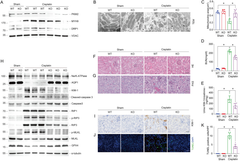
Fig. 5. Deletion of PKM2 in tubular epithelial cells inhibits mitochondrial fragmentation and alleviates cisplatin-induced AKI in vivo.
A Western blots result of mitochondrial PKM2, MYH9, and DRP1 expression in tubules from tubular-specific Pkm2 knockout mice (KO) compared to wild-type mice (WT) 1 day after cisplatin injury. B, C Representative images of Transmissionelectron microscopy (TEM) and quantification of mean mitochondrial area (μm2) in proximal tubular epithelial cells from WT mice and KO mice 3 days after cisplatin injury. scale bar = 500 nm. *P < 0.05, n = 6–7 per group. D, E The BUN level or urinary KIM-1 to creatinine ratio of WT mice and KO mice 3 days after cisplatin injury. *P < 0.05, n = 6–7 per group. F, G Representative images of H&E and PAS staining of kidney tissues from WT mice and KO mice 3 days after cisplatin injury. scale bar = 50 μm. H Western blot analysis of Na/K-ATPase, AQP1, KIM-1, cleaved-caspase3, caspase3, RIP1, p-RIP3, RIP3, p-MLKL, MLKL, and GPX4 expression in kidney tissues from WT mice and KO mice 3 days after cisplatin injury. I, J Representative images of immunohistochemical and immunofluorescent staining for KIM-1, TUNEL in kidney tissues from WT mice and KO mice 3 days after cisplatin injury. scale bar = 50 μm. (K) Quantitative analysis of TUNEL positive cells among groups as indicated. scale bar = 50 μm. *P < 0.05, n = 6–7 per group.

