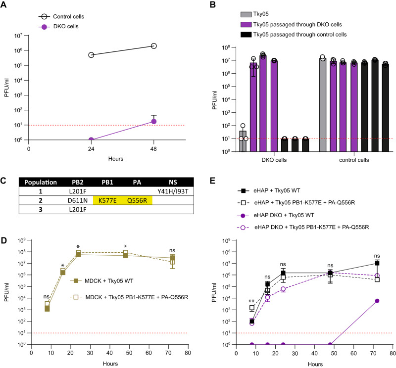
Fig. 1. An influenza A virus evolved to grow in human cells lacking ANP32A and ANP32B proteins.
A Tky05 was inoculated on 12-well plates of eHAP (control) and DKO cells at an MOI of 0.0005. Samples were titred on MDCK cells via plaque assay at 24 and 48 h post infection. Data are presented as mean values +/− SD. B Following two passages through either DKO cells or WT eHAP cells (control), 1000 PFU from each population as well as the ancestor (Tky05) were inoculated onto six-well plates of DKO and eHAP cells. After 48 h, viruses were titred on MDCK cells via plaque assay. n = 3 wells, data are presented as mean values +/− SD. C Mutations from populations passaged through DKO cells were discovered using Sanger sequencing. Highlighted mutations were found in the plaque-purified virus, which was used for future experiments. Growth curves of Tky05 compared to plaque-purified virus containing PB1 K577E, PA Q556R on MDCK (D) or eHAP (E) cells. 12-well plates were infected at MOI 0.001 (D) or 0.0005 (E), and viral titres were measured on MDCK cells at 8, 16, 24, 48 and 72 h. n = 3 wells, data are presented as mean values +/− SD. Statistical significance was determined by multiple t-test of log-transformed data, *P < 0.05, **P < 0.01, ***P < 0.001, ****P < 0.0001, (1D; 8 hpi p = 0.238, 16 hpi p = 0.206, 24 hpi p = 0.035, 48 hpi p = 0.017, 72 hpi p = 0.339. 1E; 8 hpi p = 0.003, 16 hpi p = 0.101, 24 hpi p = 0.802, 48 hpi p = 0.600, 72 hpi p = 0.066) Source data are provided as a Source Data file.

