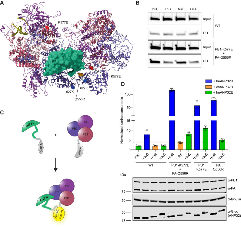
Fig. 4. PB1 K577E and PA Q556R increase binding to huANP32E.
A Asymmetric dimer of influenza polymerase (PDB: 6XZP) and ANP32 (green) showing PB2 627K in blue, PB1 K577E in red and PA Q556R in yellow. B Co-immunoprecipitation assays were conducted from 10 cm dishes of eHAP TKO cells transfected with pCAGGS Tky05 PB2 (5 µg), Tky05 PB1 or K577E (5 µg), Tky05 PA or Q556R (5 µg) and huANP32B-FLAG/chANP32B-FLAG/huANP32E-FLAG (5 µg). Twenty-four hours after transfection, cells were lysed, FLAG-tagged proteins were immunoprecipitated, and co-precipitation of PB2 was detected using immunoblotting. Data presented are representative of n = 3 biological repeats. C Schematic showing the reconstitution of Gaussia luciferase activity from the interaction of the C-terminus of Gaussia luciferase fused to ANP32 with the N-terminus of Gaussia luciferase fused to PB1. D Split luciferase assay measuring the interaction between influenza polymerase combinations Tky05 WT, PB1 K577E + PA Q556R, PB1-K577E and PA-Q556R (formed using PB1-Gluc1 or PB1-K577E-Gluc1 fusions) and either huANP32B-Gluc2, chANP32B-Gluc2 or huANP32E-Gluc2. Data presented are representative of n = 3 biological repeats each conducted with n = 3 wells, presented as mean values +/− SD. Accompanying Western blot showing expression of PA/Q556R, PB1/K577E-Gluc1, tubulin and ANP32-Gluc2. Source data are provided as a Source Data file.

