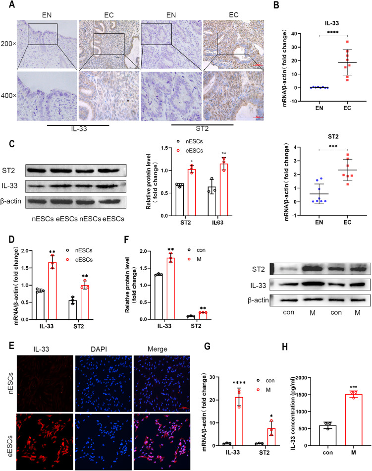
Fig. 1. IL-33 from co-cultured macrophages activates ST2 in eESCs.
A Representative immunohistochemical images staining with IL-33 and ST2 in normal endometrial tissue (EN) and ectopic endometriosis lesion tissue (EC). (original magnification ×200 or ×400) (n = 5). B RT-qPCR was used to determine the mRNA levels of IL-33 and ST2 in EN and EC (n = 8). C Western blot was used to detect the protein levels of IL-33 and ST2 in normal endometrial stromal cells (nESCs) and ectopic endometrial stromal cells (eESCs). D RT-qPCR was used to determine the mRNA levels of IL-33 and ST2 in nESCs and eESCs. E Representative immunofluorescence (IF) images of IL-33 (red) in nESCs and eESCs, Nuclei were stained with DAPI (blue). (original magnification ×200). F Western blot was used to detect the protein levels of IL-33 and ST2 in eESCs with or without macrophages co-culture treatment. G RT-qPCR was used to determine the mRNA levels of IL-33 and ST2 in eESCs with or without macrophages co-culture treatment. H ELISA analysis of IL-33 concentration in eESCs culture medium with or without macrophages co-culture treatment. Data are presented as the mean ± SD, n = 3 independent experiments. Statistical analysis was performed using Student’s t test. ****p < 0.0001, ***p < 0.001, **p < 0.01, *p < 0.05. IL-33 interleukin-33, ST2 interleukin receptor-like 1 (IL1R-L1), M macrophages, con control.

