
Fig. 7.
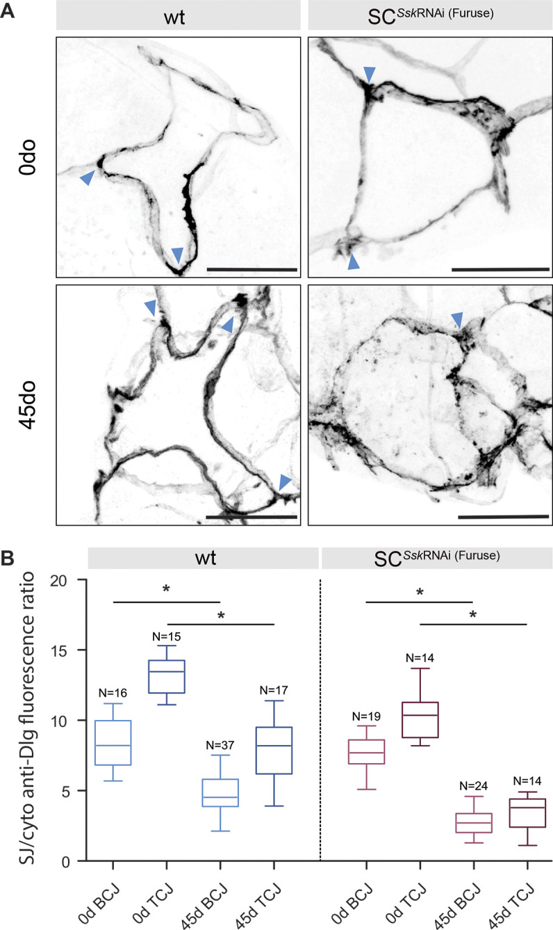
SC-specific depletion of Ssk impairment advances age-related changes in junctional protein localisation. (A) Distribution of Dlg in adult wild-type (wt) and SCSskRNAi SCs at 0 and 45 days post eclosion. Tricellular junctions are indicated by arrowheads. Scale bars: 20 μm. (B) Graphical representation of the ratio of Dlg fluorescence signal at bicellular (BCJ) and tricellular (TCJ) junctions compared with cytoplasmic signals in adult wt and SCSskRNAi SCs at 0 and 45 days post eclosion. Boxes show the 25–75th percentiles, whiskers show the minimum and maximum values, and the median is marked with a line. *P<0.0001, two-tailed unpaired Student's t-test.