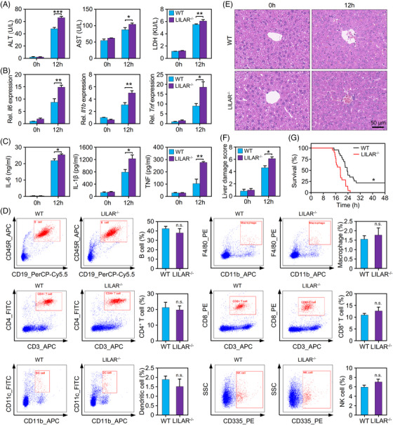
FIGURE 2.

Deficiency of LILAR aggravates liver injury in endotoxemic mice induced by lipopolysaccharide (LPS). (A) Quantification of alanine aminotransferase (ALT), aspartate aminotransferase (AST), and lactate dehydrogenase (LDH) in the serum of wild‐type (WT) and LILAR knockout (KO) (LILAR −/−) mice treated without or with LPS for 12 h. n = 3/group. (B) Quantitation of Il6, Il1b, and Tnf mRNA expression in the liver tissues of WT and LILAR −/− mice treated without or with LPS for 12 h by quantitative real‐time polymerase chain reaction (qRT‐PCR). n = 3/group. (C) Protein quantitation of interleukin‐6 (IL‐6), IL‐1β, and tumor necrosis factor (TNF) in the serum of WT and LILAR −/− mice treated without or with LPS for 12 h. n = 3/group. (D) Influence assessment of LILAR deficiency on immune system development. A single‐cell suspension was obtained from the spleens of WT and LILAR −/− mice. Flow cytometric analysis of splenic immune cells was performed with specific antibodies as indicated, and cell populations were gated based on the area and aspect ratio. n = 3/group. (E and F) Histopathological examination of livers from WT and LILAR −/− mice treated without or with LPS for 12 h. Hematoxylin and eosin (H&E) staining (E) and liver tissue damage score assessment (F) were performed as described in the methods. n = 6/group. (G) Survival curves of WT and LILAR −/− mice (n = 20/group) treated with LPS. Statistical analysis was performed using the log‐rank (Mantel–Cox) test. * p < 0.05; ** p < 0.01; *** p < 0.001; n.s., no significance.
