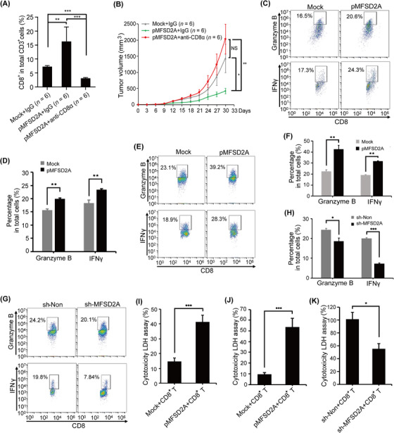
FIGURE 3.

MFSD2A expression in GC cells enhances CD8+ T cell activation. (A) Flow cytometry analysis of the proportion of tumor infiltrating CD8+ T cells in tumor tissue from MFSD2A‐overexpressed MFC cells bearing 615 mice or control mice administrated with CD8α antibody or control IgG. (B) Depletion of CD8+ T cells by CD8α antibody rescued the inhibitory effect of MFSD2A overexpression on GC growth. (C‐D) Tumor‐infiltrating CD8+ T cells were co‐cultured with MFSD2A‐overexpressed or control MFC cells for 24 h, and the expression of Granzyme B and IFNγ produced by T cells was analyzed by flow cytometry. (E‐H) CD8+ T cells from human PBMCs were co‐cultured with MFSD2A‐overexpressed or control MGC803 cells (E and F), MFSD2A‐silenced MGC803 cells or control cells (G and H) for 24 h. The expression of granzyme B and IFNγ produced by T cells was analyzed by flow cytometry. (I‐K) The cytotoxicity of CD8+ T cells from MFSD2A‐overexpressed MFC tumor (I) and of PBMCs (J and K) after co‐cultured with MFSD2A‐overexpressed MFC or MGC803 cells or control cells (I and J) or with MFSD2A‐silenced MGC803 cells or control MGC803 cells (K) for 24 h was assayed by LDH analysis. All values are presented as the mean ± SEM. * P < 0.05, ** P < 0.01, *** P < 0.001, NS, not significant. Abbreviations: MFSD2A, Major Facilitator Superfamily Domain Containing 2A; IFNγ, Interferon‐gamma; LDH, lactate dehydrogenase; PBMCs, peripheral blood mononuclear cells; GC, gastric cancer; SEM, standard error of the mean.
