Fig. 2.

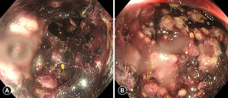
Endoscopic image showing ischemic colitis. (A) Sigmoidoscopy showing multiple various-sized bullae with hemorrhage from the rectum to the distal transverse colon. (B) Focal yellowish pseudomembranes can been seen among the bullae.

Endoscopic image showing ischemic colitis. (A) Sigmoidoscopy showing multiple various-sized bullae with hemorrhage from the rectum to the distal transverse colon. (B) Focal yellowish pseudomembranes can been seen among the bullae.