Figure 3.
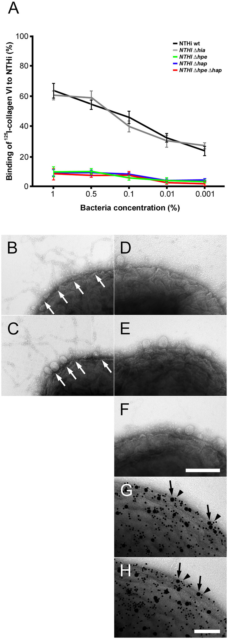
Targeting of NTHi surface adhesins PE and Hap by collagen VI VWA domains. (A) Titration of bacterial solutions with radiolabeled collagen VI microfibrils. Serial dilutions of bacteria were used: 1% (2 × 10⁹ cfu/ml), 0.5% (1 × 10⁹ cfu/ml), 0.1% (2 × 10⁸ cfu/ml), 0.01% (2 × 10⁷ cfu/ml), and 0.001% (2 × 10⁶ cfu/ml). Wild type bacteria are compared to isogenic mutants as indicated. (B–F) negative staining and transmission electron microscopy of collagen VI networks bound to the bacterial surface. Wild type (B) and Δhia (C) bacteria interact with collagen VI (arrows) as opposed to Δhpe (D), Δhap (E), and ΔhpeΔhap (F). PE (G) and Hap (H) are frequently colocalized with collagen VI on the bacterial surface as visualized by antibodies conjugated with 5 nm (PE and Hap, arrowheads) and 10 nm (collagen VI, arrows) colloidal gold, respectively. The scale bars represent 200 nm (B–F) and 100 nm (G, H).

