
Fig. 2. Loss of the C1P-cPLA2α interaction induces peritoneal neutrophilia in E. coli infection, improves survival outcomes in sepsis, and enhances the closure rate of pressure ulcers.
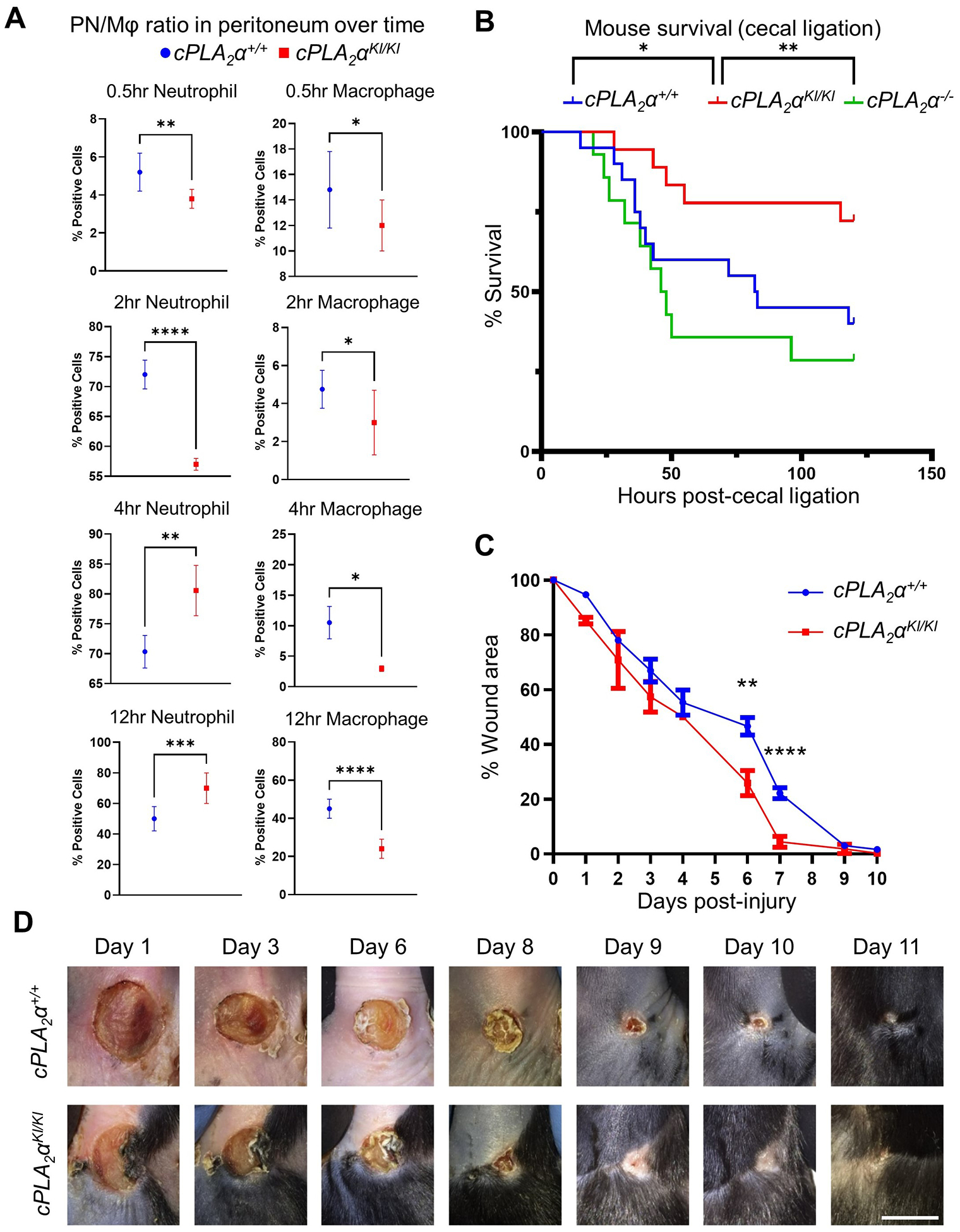
(A) Quantification of neutrophils and macrophages in peritoneal cells isolated from cPLA2α+/+, cPLA2αKI/KI, and cPLA2α−/− mice after intraperitoneal (IP) injection of E. coli. At the indicated times, peritoneal cells were extracted, sorted for neutrophil and macrophage markers (LY6G++ high and F4/80++ high, respectively), and quantified by FACS analysis. Data are displayed as mean ± SD and were statistically analyzed by unpaired t-test with Welch’s correction; *p < 0.05, **p < 0.01, ***p < 0.001, ****p < 0.0001. n=4–8 mice per genotype for each noted time point and repeated on at least two separate occasions. (B) Survival of cPLA2α+/+, cPLA2αKI/KI, and cPLA2α−/− mice subjected to cecal ligation and puncture to induce sepsis. The Kaplan-Meier method was used to estimate survival and statistically analyzed with respect to survival using the Log-rank Mantel-Cox test (cPLA2αKI/KI vs cPLA2α−/− **p = 0.0034, cPLA2αKI/KI vs cPLA2α+/+ *p = 0.0430, n=15–20 mice per genotype repeated on more than two occasions). (C and D) Quantification (C) and representative images (D) of wound closure in cPLA2α+/+ and cPLA2αKI/KI mice subjected to the Stadler model of pressure ulcers by ischemia and reperfusion. At the indicated times, wound area was assessed by change in area versus day 0. Boxplots are displayed. Data analyzed using Repeated Measures ANOVA; **p < 0.01, ****p<0.0001); n= 5–8 mice per genotype repeated on more than two occasions). Scale bar, 5 mm.