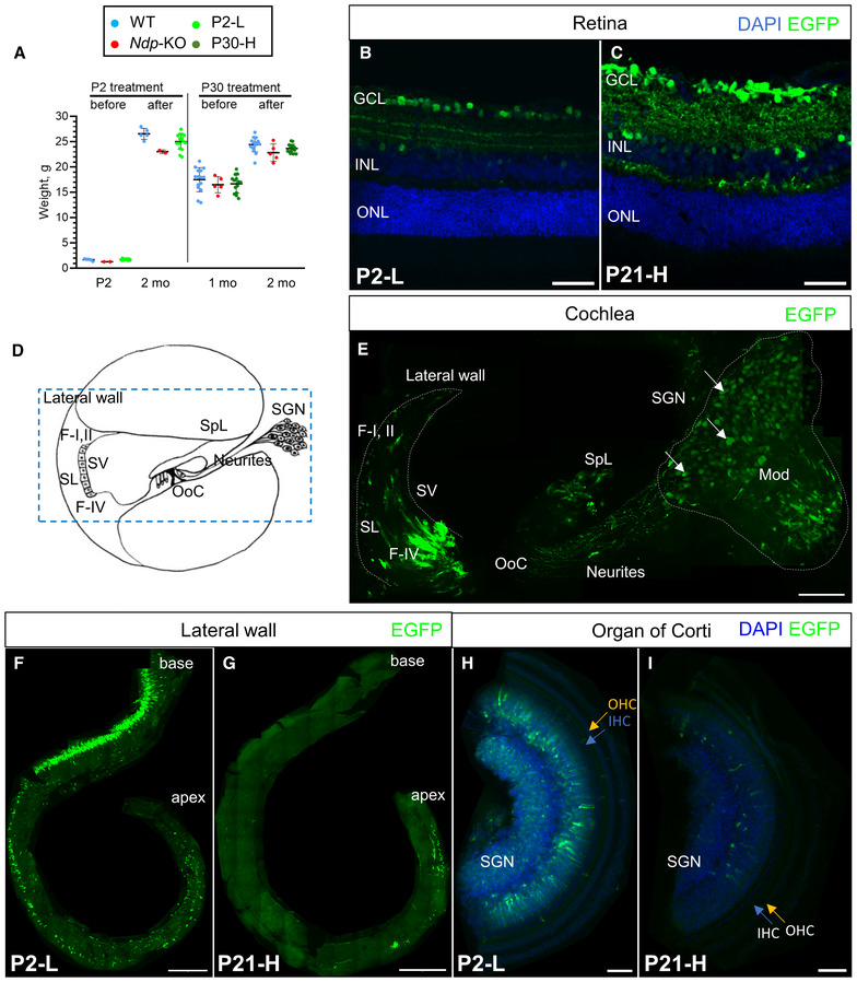
Figure 2. Expression of the construct in eye and ear at 2 months.

-
AWeights of male treated mice and age‐matched litter mate controls before and after AAV9.NDP administration. Data are shown as mean ± SD. Animal numbers: P2‐L group, n (WT) = 5, n (Ndp‐KO) = 2, n (P2‐L) = 16; P30‐H group, n (WT) = 15, n (Ndp‐KO) = 5, n (P30‐H) = 14.
-
B, CAAV9.NDP transduction at 2 months in P2 and P21 treated retinal sections: (B) P2‐L group, n = 4; (C) P21‐H group, n = 4. Staining: anti‐GFP antibody (EGFP, green). GCL – ganglion cell layer, ONL – outer nuclear layer, INL – inner nuclear layer. Scale bar 50 μm.
-
DA schematic of the axial cross‐section of one turn of the cochlea. SL, spiral ligament; SV, stria vascularis; SGN, spiral ganglion neurons; OoC, organ of Corti; SpL, spiral limbus; F‐I, II, type I and II fibrocytes; F‐IV, type IV fibrocytes. Blue dashed rectangle outlines region, shown in (E). Blue solid rectangle outlines spiral ganglia region.
-
E–IAAV9.NDP transduction in cochlea: anti‐EGFP antibody (green) and DAPI (blue). Transduction in P2‐treated mouse cochlea at 2 months, cross‐section corresponding to the dashed outline in (D) (E). Transduction of the lateral wall of the cochlea in wholemounts at 2 months after treatment at P2 (F) and P21 (G). Scale bar: 50 μm. Transduction of the organ of Corti in wholemounts at 2 months after treatment at P2 (H) and P30 (I). Scale bar: 100 μm. Appendix Fig S4 shows extended views of cochlear transduction.
Source data are available online for this figure.
