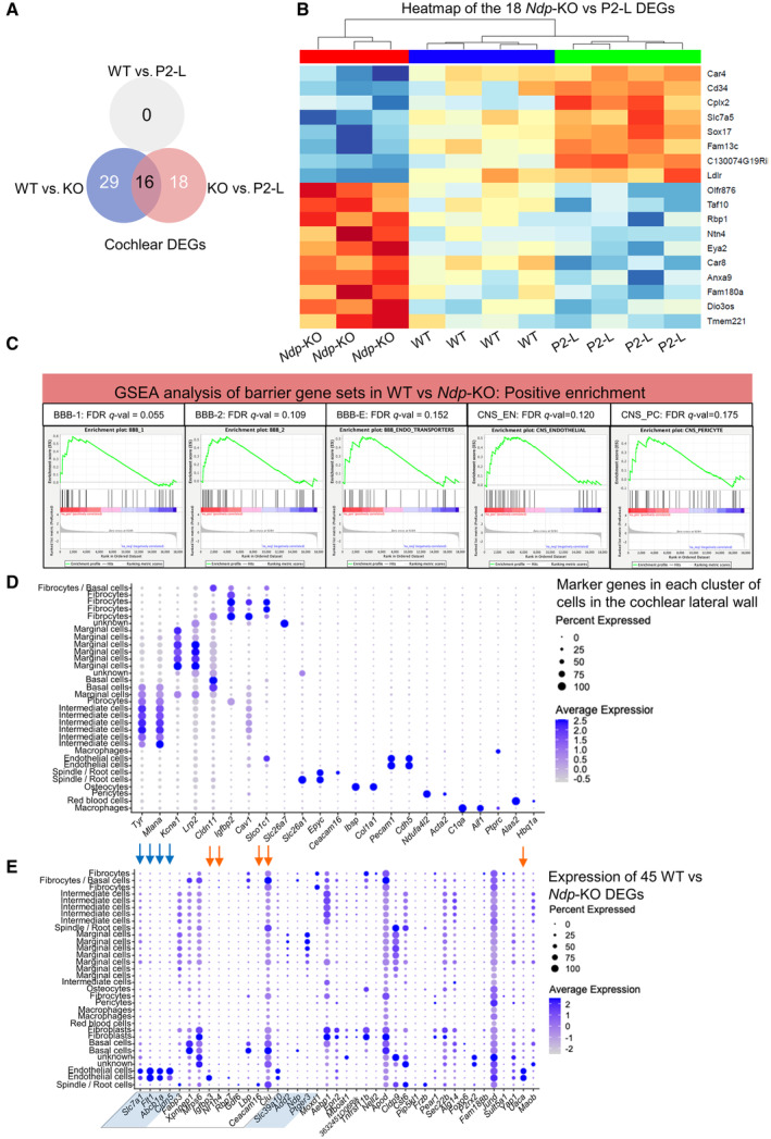
Figure EV4. Differential gene expression in the cochlea by RNA sequencing and qRT‐PCR.

- Venn diagram showing overlap between Ndp‐KO versus WT DEGs and Ndp‐KO versus P2‐L DEGs.
- Heatmap showing levels of expression of the 18 pathology‐related DEGs identified between Ndp‐KO (red) versus P2‐L (blue) in WT, Ndp‐KO and P2‐L treated cochleas (green). Note that these genes showed trends of differential expression between Ndp‐KO and WT cochleas that did not reach significance. No genes were found to be significantly differentially expressed between WT and P2‐L cochleas, indicating rescue by the treatment. In heat map, red indicates upregulated and blue indicates downregulated gene expression in Ndp‐KO.
- Gene set enrichment analyses of differentially expressed genes between WT and Ndp‐KO using gene sets were previously defined by transcriptome profiling of FACS sorted CNS vs peripheral endothelial cells (Daneman et al, 2010) and previously used to assess transcriptomes of WT and Ndp‐KO retinas (Zhou et al, 2014). Gene sets characterising barrier vasculature, BBB1 and 2, BBB endothelial transporters, CNS endothelial and CNS pericyte were significantly positively correlated (FDR < 0.25) with the WT genotype.
- Dot plot using scRNA seq data of the adult mouse cochlear lateral wall from GEO database: accession numbers GSM5124299, GSM5124300, GSM5124301, and GSM5124302. Gene markers used to distinguish 30 cell type clusters in the UMAP were as previously reported (Gu et al, 2020; Bryant et al, 2022).
- Dot plot showing expression of the 45 DEGs identified in WT versus Ndp‐KO analysis in the 30 cell type clusters identified in the adult mouse cochlear lateral wall at the single cell level. Blue arrows indicate endothelial cell genes (Cldn5, Abcb1a and Flt1) downregulated in Ndp‐KO, and orange arrows indicate upregulated genes in Ndp‐KO. Box indicates genes significantly different in the P2‐L versus Ndp‐KO comparison. Shaded DEGs are downregulated in the Ndp‐KO cochlea. Note expression of some DEGs across several different cell type clusters.
