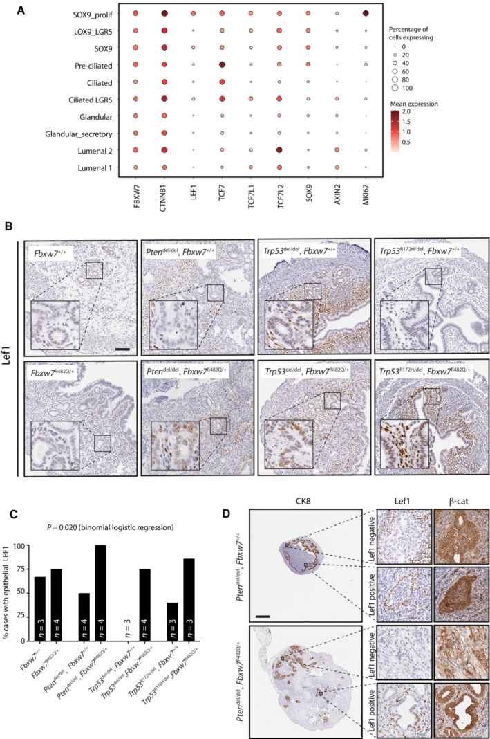
Figure 6. LEF1 is highly expressed in proliferative endometrial stem cells and dysregulated by Fbxw7 hotspot mutation.

- Dot plot showing log2‐transformed expression of FBXW7, LEF1 and other wnt pathway genes in endometrial epithelial cell subsets defined by single cell RNAseq (scRNAseq) in Garcia‐Alonso et al (2021).
- Representative LEF1 immunohistochemistry in GEMM uteri from mice of indicated genotypes at 8 weeks age. For each of the four columns (i.e genotypes), upper and lower images are taken from littermate females housed in the same cage. Images are representative of 3–4 mice as shown in (C). Scale bar indicates 100 μm.
- Percentage of mice with epithelial LEF1 expression in endometria at 8 weeks age according to genotype. P value was derived from binomial logistic regression with epithelial LEF1 expression as the dependent variable.
- Immunohistochemistry for LEF1 and β‐catenin from endometrial carcinomas in Pten del ± Fbxw7 mut mice. High power images show representative staining in LEF1 negative and positive glands. Scale bar indicates 1,000 μm.
Source data are available online for this figure.
