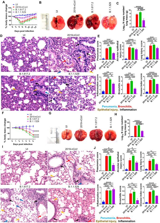
Fig. 2.
Pathological manifestations of Omicron (B.1.1.529) infection in Syrian hamster and hACE2.Tg mice. Pathological manifestations of intranasal Omicron (B.1.1.529) infection was evaluated and compared with ancestral Wuhan (nCoV-2019) and Delta (B.1.617.2) strain infection or uninfected (UI) in Syrian hamster and hACE2.Tg mice. The changes in body mass was plotted as percentage of the day 0 body mass till day 14 or 6 post infection, respectively, for (A) hamster and (F) hACE2.Tg mice. The lungs of the sacrificed animals were harvested and images and thereafter, viral load and histopathology of the lungs were studied. B and G Shows representative lungs from individual groups at 4 dpi and 6 dpi from hamster and hACE2.Tg mice, respectively. C and H Viral load of the lungs at 4dpi and 6 dpi from hamster and hACE2.Tg mice, respectively. D–E and I–J Histopathological assessment of the HE stained lungs were carried out by trained pathologist by blinded scoring on the scale of 0–5 (where 0 described no feature and 5 described highest pathological feature) of the lungs at 4 dpi and 6 dpi from hamster and hACE2.Tg mice, respectively. Each experiment was carried out with n = 5 animals and replicated 3 times independently. One-way ANOVA using non-parametric Kruskal–Wallis test for multiple comparison. ns = non-significant, *P < 0.05, **P < 0.01, ***P < 0.001, ****P < 0.0001

