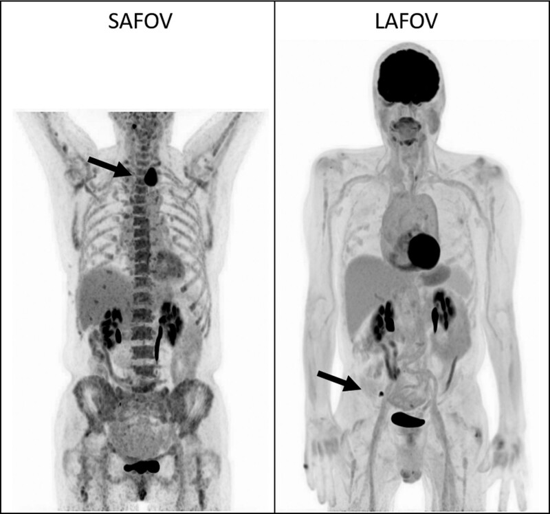
Fig. 5.
Example maximum intensity projection (MIP) for random patients undergoing lymphoma assessment on a SAFOV (left) and LAFOV (right) system with hypermetabolic disease in both patients highlighted by the solid arrows. The LAFOV images exhibit improved axial coverage and lower noise compared to the SAFOV (SUV window for both images 0–6). LAFOV, long-axial field-of-view; SAFOV, short-axial field-of-view.

