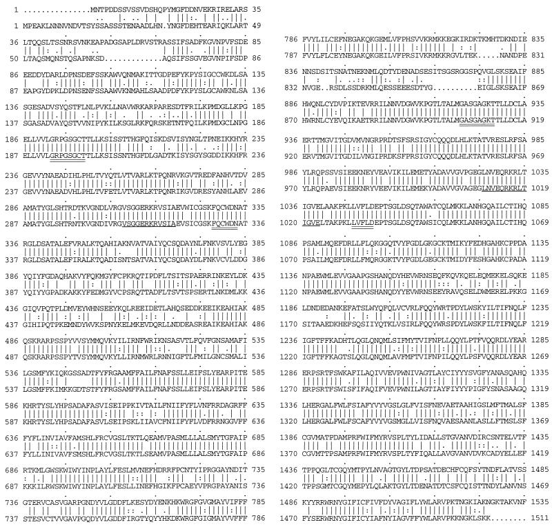
FIG. 4.
Comparison of the amino acid sequences of PDH1 and PDR5. For each pair of sequences, the top sequence is PDH1 and the bottom one is PDR5. Lines between the sequences indicate perfect matches of amino acids. Periods and colons between the sequences show similarity based on the Dayhoff table (30a) as described elsewhere (7a). There was 72.5% identity between the two sequences, with two gaps. Putative Walker A and B sites (double underlines) and putative ATP-binding sites (single underline) are indicated.

