Figure 4.

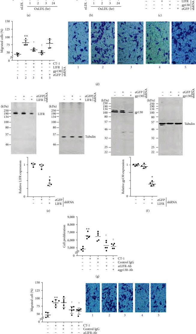
OxLDL stimulates SMC migration and proliferation via CT-1 induction. (a, b) OxLDL stimulates CT-1 mRNA expression and secretion. Quiescent SMCs treated with OxLDL for the indicated periods were analyzed for CT-1 mRNA expression by RT-qPCR and its secreted levels in equal amounts of culture supernatants by ELISA. nLDL served as a control. (c–f) CT-1 stimulates SMC migration and proliferation via LIFR and gp130. SMCs were transduced with validated lentiviral LIFR or gp130 shRNA, made quiescent, and exposed to CT-1. Cell proliferation after 48 hr (c) and migration after 18 hr (d) were analyzed by CyQUANT GR dye assay and Boyden chamber assay, respectively. The inset in (d) shows representative images of Matrigel™ transwell invasion. Knockdown of LIFR and gp130 was confirmed by western blotting (e, f), and summarized semiquantification of the intensity of immunoreactive bands is shown in the lower panels. (g, h), Preincubation with neutralizing anti-LIFR or anti-gp130 antibodies blunt CT-1-induced SMC proliferation and migration. The inset in (h) shows representative images of Matrigel™ transwell invasion. (a–d, g, h) ∗0.05, ∗∗P < 0.01 versus nLDL (n = 4 or 5); (e, f) ∗P < 0.05 versus eGFP shRNA (n = 3).
