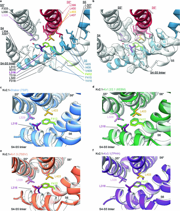
Extended Data Fig. 8. Additional hydrophobic residues interacting with the hydrophobic coupling nexus in Kv2.1 and structural alignment of Kv2.1 with other Kv channels.
a) View of the hydrophobic coupling nexus residues highlighted with the side chains depicted as sticks with F412 green, L316 purple, L329 light purple and L403 yellow, with additional hydrophobic residues depicted as sticks coloured based on the helix in which they are located. b) Same view and model as in panel a but also showing cryo-EM density for hydrophobic side chains. c-f) Close-up view of the hydrophobic coupling nexus residues highlighted with the side chains depicted as sticks with Kv2.1 residues labeled for Kv2.1 (white) and c) Shaker-IR (blue), d) Kv1.2/2.1 paddle chimera (green), e) Kv1.3 (orange) and f) Kv3.1 (purple).

