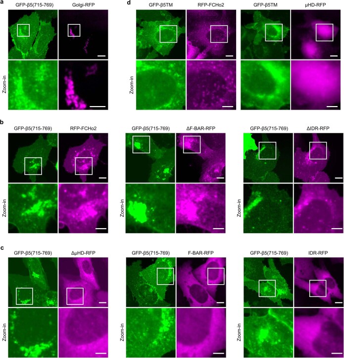
Extended Data Fig. 8. GFP-β5(715-769) induces dramatic redistribution of μHD-containing FCHo2 variants to the plasma membrane and the Golgi apparatus.
a, GFP-β5(715-769) is located on the plasma membrane with some accumulations around the perinuclear region that colocalizes with Golgi apparatus marker Golgi-RFP. Scale: full-size, 10 µm; insets, 5 µm. b, When co-expressed with GFP-β5(715-769), three μHD domain-containing FCHo2 variants (RFP-FCHo2, FCHo2_ΔF-BAR-RFP, and FCHo2_ΔIDR-RFP) are redistributed to the plasma membrane and colocalize with GFP-β5(715-769) in the perinuclear region. Scale: full-size, 10 µm; insets, 5 µm. c, When co-expressed with GFP-β5(715-769), three FCHo2 variants that don’t contain μHD domain (FCHo2_ΔµHD-RFP, FCHo2_F-BAR-RFP and FCHo2_IDR-RFP) are highly diffusive in the cytosol. d, When co-expressed with the negative control GFP-β5TM, RFP-FCHo2 shows cytosolic diffusive pattern with small puncta (left) and FCHo2_µHD is highly cytosolic (right and in Fig. 4l). Scale: full-size, 10 µm; insets, 5 µm.

