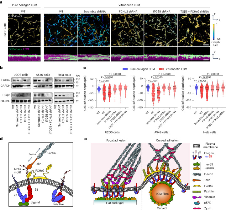
Fig. 6. Curved adhesions facilitate cell migration in 3D ECMs.
a, Representative images of U2OS cells expressing GFP–CaaX in 3D matrices after 72 h of culture. Cells are colour-coded according to z depth in x–y projections. Cells are coloured in green and merged with ECM in magenta in x–z projections. WT cells infiltrated into matrices made of vitronectin fibres, but not matrices made of pure collagen fibres. The shRNAs of FCHo2, ITGβ5 or both, but not scramble shRNA, inhibited the cell infiltrations. Scale bar, 100 µm. b, Western blots show that shRNAs are able to effectively reduce the expression of FCHo2, ITGβ5 or both in U2OS, A549 and Hela cells. These results support the knockdown efficiencies supplied by the company (Supplementary Table 3). c, Quantification of the cell infiltration depth for U2OS, A549 and Hela cells in 3D pure collagen matrices or 3D vitronectin matrices with the knockdown of FCHo2, ITGβ5 or both. Left to right, n = 244, 509, 348, 279, 251 and 211 U2OS cells, n = 164, 134, 100, 238, 92 and 177 A549 cells and n = 142, 236, 277,189, 241 and 204 Hela cells, from 2 independent experiments. Medians (lines) and quartiles (dotted lines) are shown. P values calculated using Kruskal–Wallis test with Dunn’s multiple comparison. d, Illustration of ITGβ5 interacting with FCHo2 in curved adhesions. e, Schematic comparison of focal adhesion and curved adhesion architectures. Note that the models only depict proteins investigated in this work. Source numerical data and unprocessed blots are available in the source data.

