Fig. 1.

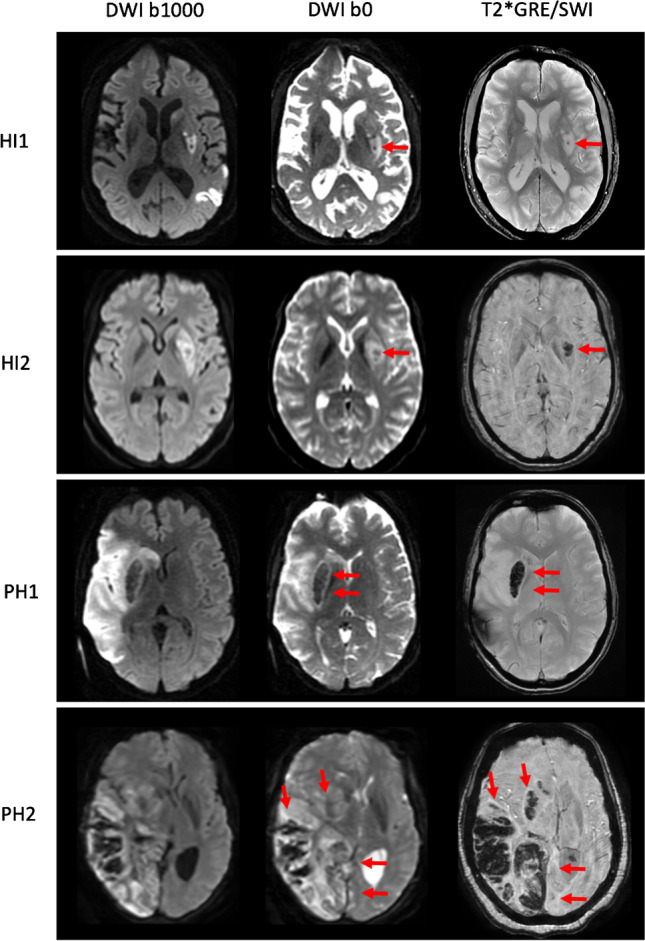
Detection of intracranial hemorrhage (ICH) on diffusion-weighted imaging (DWI) b0 and T2*GRE/SWI. Axial slices of four examinations are displayed from top to bottom showing HI1, HI2, PH1, and PH2 types of ICH indicated by the red arrows

Detection of intracranial hemorrhage (ICH) on diffusion-weighted imaging (DWI) b0 and T2*GRE/SWI. Axial slices of four examinations are displayed from top to bottom showing HI1, HI2, PH1, and PH2 types of ICH indicated by the red arrows