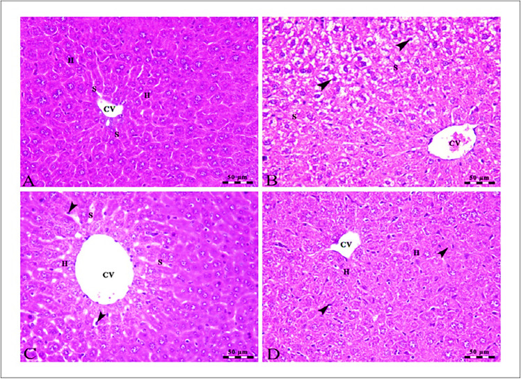
Fig. 4.
A Photomicrographs of the liver of the control group showing intact polyhedral-shaped hepatocytes (H) with a rounded centrally located nucleus arranged in a cord-like pattern and separated by narrow blood sinusoids (S) which radiates from the intact central vein (CV). B The centro lobular area of the liver of the SOR-treated group showing mild swelling and vacuolar degeneration of some hepatocytes at the periphery of the lobule (arrowheads) besides congestion in both central veins (CV) and hepatic sinusoids (S). C The liver of the CLT-treated group showing mild micro steatosis in the hepatocytes (H), mild dilated central vein (CV), and blood sinusoids (S) in addition to the presence of Kupffer cells (arrowheads). D The centro lobular area of the liver of the SOR + CLT-treated group showing central vein (CV), intact hepatocytes arranged in cords (H), and the presence of Kupffer cells activity (arrowheads). Stain H&E. Scale bars = 50 µm

