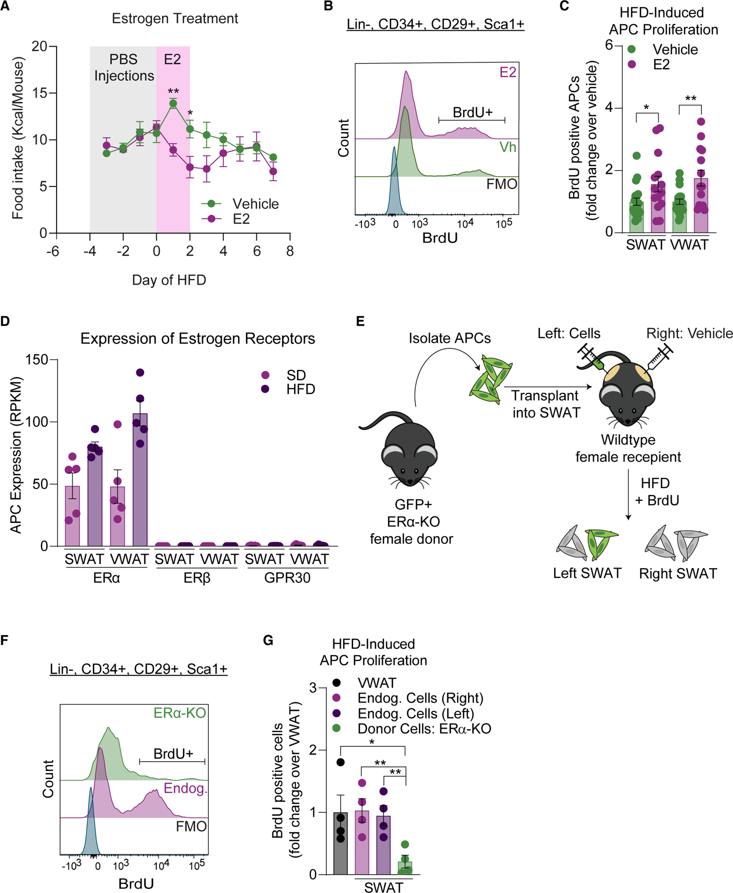
Figure 3. Estradiol drives female APC proliferation in an ERα-dependent manner.

(A) Food intake (cage average) during vehicle or E2 treatment (days 0–2 of an HFD) in female mice (n = 14–19 mice per group).
(B) Representative BrdU histograms in APCs from estradiol treatment experiment including BrdU FMO.
(C) APC proliferation after 1 week of HFD feeding in vehicle or E2 treated female mice.
(D) Expression of estrogen receptors in female APCs from RNA-seq data (n = 5 samples per group, 3 mice pooled per sample).
(E) Schematic of APC transplantation assay into female SWAT.
(F) Representative BrdU histograms in APCs from APC-ERαKO transplant experiment including BrdU FMO.
(G) APC proliferation of transplanted ERα-KO and endogenous APCs after 1 week of HFD feeding in females (n = 4 mice per group).
Statistical significance was determined by one-way ANOVA with Šidák’s tests for (A) and unpaired t tests for (C) and (F). Error bars represent mean ± SEM. ns, not significant, *p < 0.05, **p < 0.01. APCs, adipocyte precursor cells; VWAT, perigonadal fat; SWAT, inguinal subcutaneous fat; BrdU, bromodeoxyuridine; E2, 17β-estradiol; Vh, vehicle; SD, standard diet; HFD, high-fat diet; BrdU, bromodeoxyuridine; Endog, endogenous; FMO, fluorescence minus one.
See also Figure S2.
