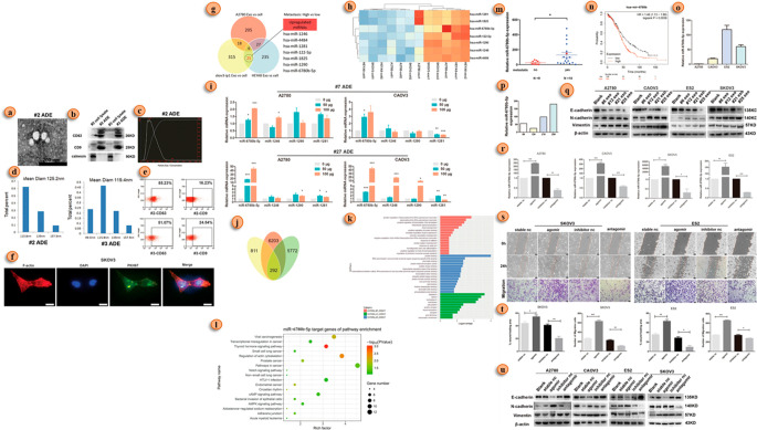
Figure 4.
Exosome miRNA-led EMT in ovarian cancer. (a) Electron microscopic view of the exosome of a patient with ovarian cancer. (b) Expression of an exosome’s marker (CD63, CD9, and calnexin) in Western blot. (c) Exosomes size analysis using a NTA (nanoparticle tracking assay). (d) Exosome size analysis via DLS (dynamic light scattering). (e) Analysis of exosome marker expression using flow cytometric analysis. (f) Exosome uptake of ovarian cancer cell analysis via immunofluorescence detection (in exosomes, PKH67 was labeled with green, and F-actin was labeled red; nuclei were labeled with DAPI). (g) miRNA analysis in three ovarian cancer cell lines. (h) Seven miRNAs’ higher expression detects in thermograms among 25 miRNAs. (i) qRT-PCR analysis of the seven most expressive miRNAs in three ovarian cancer cell lines. (j) Venn diagram of common higher expression miRNA (miR-6780b-5p). (k) Gene function annotation indicates the target gene of miR-6780b-5p. (l) KEGG analysis of the possible target genes of miR-6780b-5p. (m) qRT-PCR comparative analysis miR-6780b-5p expression. (n) Kaplan–Meier analysis of miR-6780b in pancancer analysis with a K–M plotter. (o) qRT-PCR comparative analysis of miR-6780b-5p expression in four ovarian cancer cell lines. (p) Comparative qRT-PCR analysis of miR-6780b-5p expression in four sets of clinical samples. (q) EMT marker expression in four ovarian cancer cell lines via Western blot (WB). (r) qPCR validation of the effects of the miR-6780b-5p agomir and antagomir in four cell lines. (s) Migration assay analysis after miR-6780b-5p transfection. (t) Migration assay results represented via histograms. (u) WB analysis of EMT marker expression after transfection of miR-6780b-5p. Adapted with permission from ref (83). Copyright 2021. Cell Death and Disease, Springer Nature.

