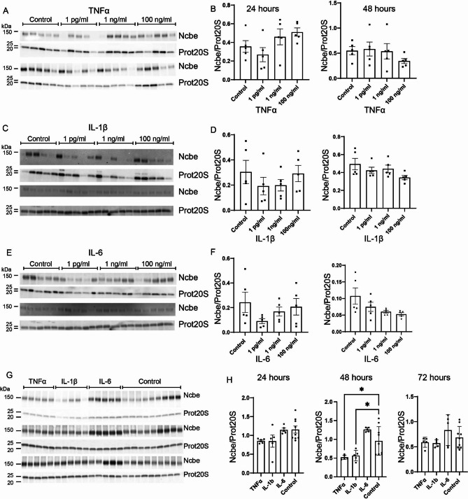
Fig. 2.
Effect of proinflammatory cytokines TNFα, IL-1β and IL-6 on CP expression of the basolateral transporter, Ncbe. Primary cultures were treated with 1 pg/mL, 1 ng/mL, or 100 ng/mL of either TNFα (A and B), IL-1β (C and D) or IL-6 (E and F) for 24 and 48 h. (G and H). Treatment with 100 ng/mL TNFα, IL-1β and IL-6 for 24, 48 and 72 h. Cells were lysed and subjected to immunoblotting for Ncbe and proteasome 20 S on the same membrane. Top two bands in A, C, E and G represent Ncbe and prot20S after 24 h treatment and lower two bands represent Ncbe and prot20S after 48 h treatment. Molecular masses (shown in kDa) are indicated on the left. The Ncbe and proteasome 20 S specific bands were quantified densiometrically. Bargraphs in B, D, F, and H show the Ncbe abundance relative to proteasome 20 S as mean ± SEM. *indicates p < 0.05

