Figure 6.
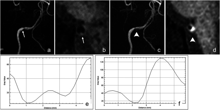
Images obtained from a 54-year-old man who presented with a sudden headache. (a) TOF-MRA at the onset shows mild focal stenosis on his right vertebral artery (arrow). (b) Z-HB imaging of the stenotic segment shows an irregular vessel wall (arrow), but no intravascular structure is confirmed based on of the observer’s eyes. (c) TOF-MRA 7 days after the onset shows intramural hyperintensity (arrowheads). (d) Z-HB imaging of the stenotic segment shows eccentric hyperintensity (arrowheads). (e) Retrospectively, the profile curve of the Z-HB imaging obtained at onset is created and analyzed. The profile curve is categorized into omega type, with a signal intensity of the intravascular structure over that of the blood flow signal of 17.74, suggesting the presence of an intimal flap in VAD. f) The profile curve of the Z-HB imaging obtained 7 days after the onset is analyzed. The profile curve is categorized into the asymmetrical type, with the signal intensity of the intravascular structure over the blood flow signal being 111.00, suggesting the presence of intramural hematoma in VAD. VAD, vertebral artery dissection; Z-HB, zoomed high-resolution black blood.

