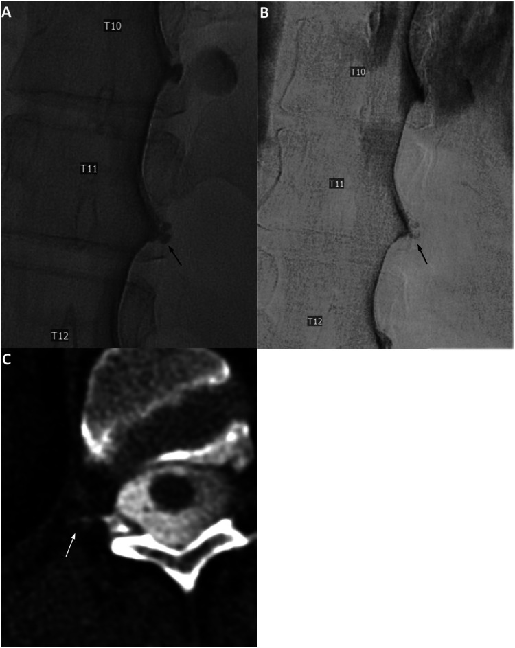
Figure 3.
Unsubtracted (a) and subtracted (b) right lateral decubitus digital subtraction myelogram images show prominent diverticulum at the right T11 level (black arrows). Subsequent CTM in the same lateral decubitus position after approximately 12 min following LDDSM (c, reformatted axial image) shows a suggestion of subtle lateral contrast extension at this level (white arrow), which was described as “indeterminant, but suspicious” for CSF-Venous fistula. Renal contrast was present (not shown). Patient underwent targeted blood patch with fibrin glue, without improvement of postural headache.

