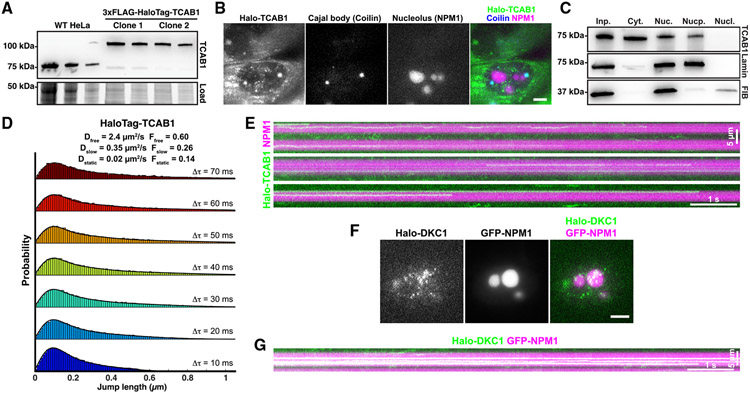
Figure 5. TCAB1 is excluded from the nucleolus.
(A) Western blot probed for TCAB1 of parental and genome-edited HeLa cells expressing HaloTag-TCAB1.
(B) Maximum intensity projection of a single-molecule imaging movie of HaloTag-TCAB1 (JFX650) in cells expressing BFP-coilin and GFP-NPM1 to mark Cajal bodies and nucleoli, respectively (scale bar, 5 μm).
(C) Western blots probed with TCAB1, fibrillarin, and lamin B1 antibodies of samples of cellular fractionation experiments (left to right: input, cytoplasm, nucleus, nucleoplasm, nucleolus) from HeLa cells.
(D) Probability density functions of the step sizes derived from nuclear HaloTag-TCAB1 trajectories and the corresponding three-state model fit using the Spot-On tool (pooled data from two replicates of two independent HaloTag-TCAB1 clones, >10 cells per replicate).
(E) Kymograph of nucleolar HaloTag-TCAB1 particles over time.
(F) Live-cell fluorescence images of single 3xFLAG-HaloTag-dyskerin particles, nucleoli marked by GFP-NPM1 (scale bar, 5 μm).
(G) Kymograph of nucleolar 3xFLAG-HaloTag-dyskerin particles over time.

