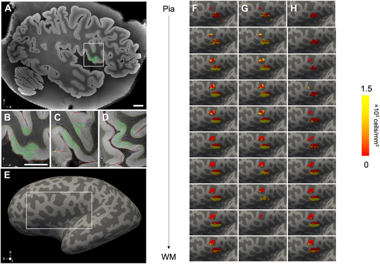
Fig. 6. Instantiation of stereological counting sites in MRI space.
(A) The stereological counting sites (green) for the NeuN markers were mapped to the MRI space by composing all spatial transformations from MRI to OCT to LSFM. (B to D) The MRI was segmented using SAMSEG, and the white matter (WM) and pial surfaces were computed using FreeSurfer; 10 equidistant intermediate surfaces were generated between the WM and pial surfaces, and counting sites were projected to their nearest vertex across all 10 surfaces (B to D: sagittal, coronal, and axial views). (E and F) The number of counted cells per vertex and total Jacobian-modulated volume of the projected sites were smoothed on the surface (three consecutive averages over the 1-ring neighbors), and their ratio yielded the density of NeuN+ neurons associated with each vertex. These densities are shown on the inflated surface. Note that only cells from layers 3 (G) and 5 (H) were counted and are included in the surface density map. Scale bars, 1 cm.

