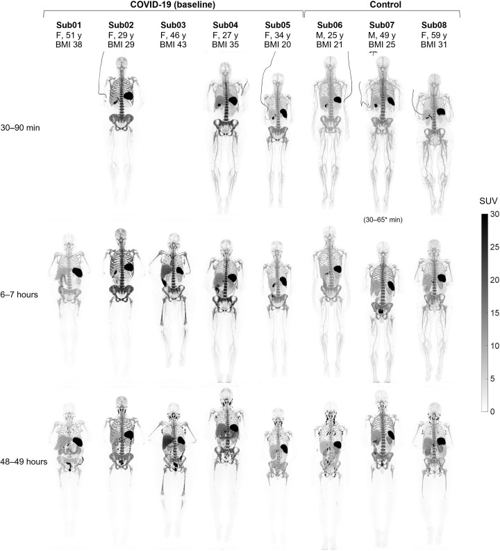
Fig. 1. Maximum intensity projection (MIP) of decay-corrected SUV images of the baseline scans.
The baseline scans of COVID-19 convalescent patients and healthy control subjects are compared at three imaging time points. Sub01 and Sub03 skipped dynamic imaging. M, male; F, female, y, years; BMI, body mass index.

