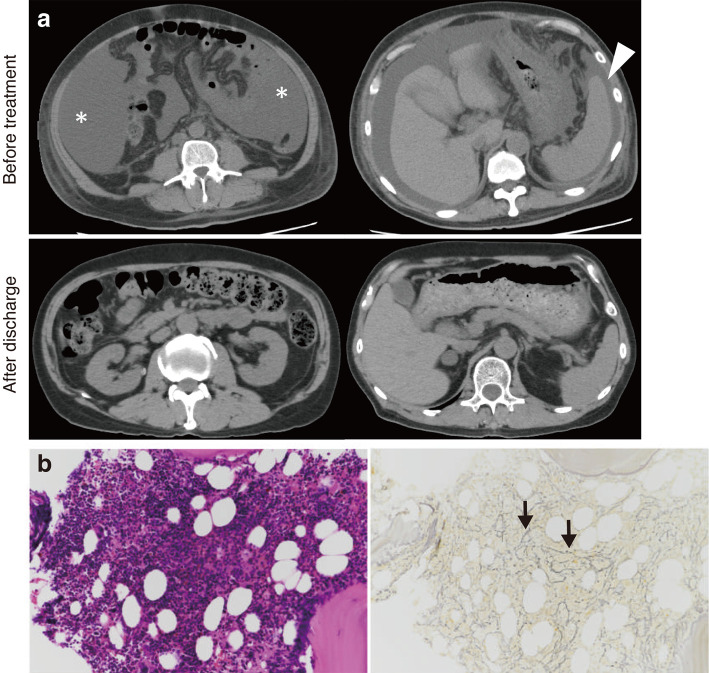
Figure 1.
(a) Plain CT shows the massive ascites (asterisk) and splenomegaly (arrowhead) before treatment. The splenomegaly and ascites improved after discharge. (b) The findings of the marrow biopsy show mildly hypercellular marrow (50% cellularity) with mild reticulin fibrosis (black arrow) (left panel: Hematoxylin and Eosin staining; right panel: silver stain, ×200).

