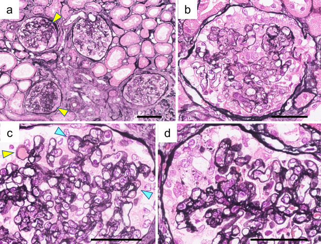
Figure 1.
Renal histological findings on light microscopy. A renal biopsy revealed the collapsing FSGS variant with concurrent endothelial damage. Periodic acid-methenamine-silver staining (PAM) revealed the following: (a) Focal segmental sclerotic lesion (yellow arrowheads). (b) Podocyte hyperplasia and endocapillary hypercellularity. (c) Hyalinosis (yellow arrowhead) and double contour (blue arrowheads). (d) Segmental collapsed capillary with podocyte hyperplasia. (a) Bars=100 μm; (b-d) Bars=50 μm.

