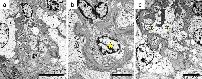
Figure 3.
Images on electron microscopy. Electron microscopy revealed podocyte injury and endothelial damage, as follows: (a) Diffuse foot process effacement, podocyte hypertrophy, and collapsed capillaries. (b) Endothelial cell swelling (star), double contour, and mesangial interposition. (c) Podocyte hypertrophy, endothelial cell swelling (star), and enlargement of the subendothelial space. (a) Bars=10 μm; (b, C) Bars=5 μm.

