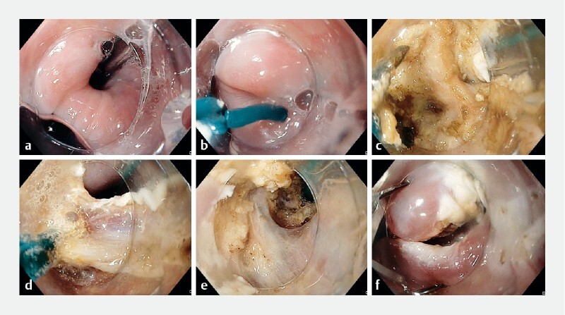
Fig. 4.

Endoscopic images during the simplified Zenker’s diverticulum peroral endoscopic myotomy (zPOEM) procedure showing: a the esophageal lumen marked with a feeding tube; b a scissor-type knife being used to cut the septum; c dissection of the mucosa to expose the cricopharyngeal muscle (CPM); d cutting of the CPM muscle; e dissection and cutting of the mucosa and the muscle until the base of the diverticulum is reached; f inspection of the base of the diverticulum, with hemostasis secured and the defect closed with clips.
