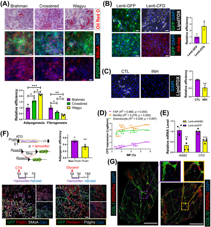
Figure 7.

The greater adipogenic potential of Wagyu FAPs is at least partially due to higher CFD expression and less robust fibrogenic programming. (A) Oil Red O staining and ICC show the adipogenic efficiency and the myofibroblast differentiation capacities of FAPs indicated by SMαA signal strength of FAPs from different breeds, respectively. The purity of FAPs was verified by PDGFRα staining. Scale bar: 50 μm (Oil Red O), 100 μm (ICC). n = 3 for each breed. (B) Brahman FAPs were transduced with Lenti‐GFP or Lenti‐CFD and induced for adipogenesis. Adipogenesis was evaluated by LipidTOX staining. The expression of GFP and CFD was verified by ICC using anti‐GFP and anti‐HA tag antibodies. n = 3 for each treatment. Scale bar: 50 μm. (C) Wagyu FAPs were treated with C3aR inhibitor (INH) or vehicle (CTL) and induced for adipogenesis. Adipogenesis was evaluated by LipidTOX staining. n = 3 for each treatment. (D) The correlation between IMF content and CFD expression in FAPs, Mo/Ma, and granulocytes. (E) Wagyu FAPs were transduced with Lenti‐shGFP or Lenti‐shNAB2. The expression levels of NAB2 and CFD were measured 2 days after transduction by realtime PCR. (F) Postn MCM/+ ;R26 eGFP mice were treated with tamoxifen from day 0 to day 3 after CTX or glycerol injections, followed by sample collection on day 7 after the CTX injection or day 14 after the glycerol injection. Muscle samples were subjected to IHC to identify Postn lineage cells (GFP+), FAPs (Pdgfrα+), myofibroblasts (Pdgfrα+;SMαA+, CTX injected), and adipocytes (perilipin‐1+, glycerol injected). The adipogenic efficiencies of Postn‐lineage FAPs [(GFP+;perilipin‐1+ cell number)/(GFP+;Pdgfrα+ cell number)] and non‐Postn‐lineage FAPs [(GFP−;perilipin‐1+ cell number)/ (GFP−;Pdgfrα+ cellnumber)] were calculated. n = 4 for each breed. Scale bar: 50 μm. (G) Representative IHC images show the locations of collagen I, collagen IV, and FAPs (PDGFRα+) in Wagyu and Brahman muscles. *P < 0.05; **P < 0.01; ***P < 0.001. COL I, collagen I; COL IV, collagen IV.
