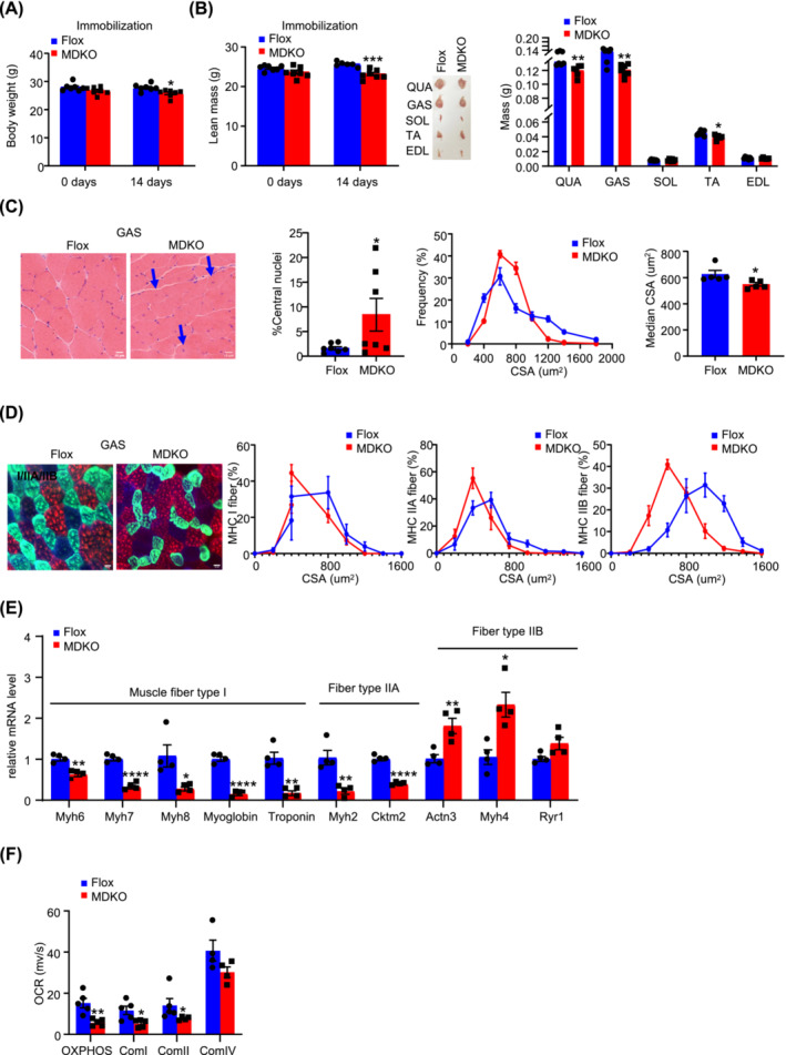
Figure 4.

DJ‐1 deletion in skeletal muscle aggravates atrophy during immobilization. (A) Body weight of male mice during immobilization at the age of 10‐week‐old (n = 6–7). (B) Lean mass, representative morphology and weight of muscles male mice during immobilization at the age of 10‐week‐old (n = 6–7). Lean mass. Representative morphology of quadriceps (QUA), GAS, soleus (SOL), TA, and extensor digitalis anterior (EDL) after immobilization. Weight of QUA, GAS, SOL, TA, and EDL after immobilization (n = 6–7). (C) Physiological consequences of GAS H&E staining in immobilized mice. Representative H&E staining of GAS (scale bars, 10 μm), the percentage of central nuclei after immobilization, CSA distribution, and median CSA in GAS of immobilized mice (n = 5). Blue arrows point to the nuclei inward migration in MDKO mice. (D) Physiological consequences of immunofluorescence staining in GAS. The different myosin heavy chain isoforms were stained in blue (MyHC‐I), green (MyHC‐IIa), and red (MyHC‐IIb) (scale bars, 20 μm). Representative immunofluorescence of muscle fibre type composition in GAS of immobilized mice and CSA distribution in GAS of immobilized mice (n = 6–7). (E) Expression of muscle fibre type related genes of GAS (n = 4). (F) Oroboros O2k respirometer oxygen flux analysis of permeabilized immobilized GAS (n = 5). Data represented the mean ± SEM. *P < 0.05, **P < 0.01, ***P < 0.001, ****P < 0.0001, a two‐tailed Student's t‐test was used for statistical analysis.
