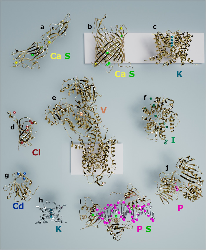
Fig. 3. Selected structures solved by native-SAD on the I23 beamline.
Protein (gold) and nucleic acids (gray) structures are shown in cartoon representation and anomalous scatterers are represented as spheres of different colors, yellow: calcium, green: sulfur, teal: potassium, red: chlorine, orange: vanadium, dark green: iodine, blue: cadmium, pink: phosphorus. The pink rectangles in the background represent the cell membrane. a Fap1. b AlgE. c NaK2K. d Streptactin. e SERCA. f TauA. g Loei River virus GP1. h RNA G-quadruplex. i IRF4-DNA. j BphA4.

